 In addition to the many exquisite rare books in the Falk Library Rare Book collection, there are a number of valuable non-literary items. As objects, they can sometimes speak to us more strongly than text, since they appeal not only to our intellect but also to our senses. Among the artifacts is the Civil War Post-Mortem Surgical Set, which was described in detail in a December 2010 HSLS Update article. Another interesting collection is the commemorative medals associated with the field of medicine. These medals consist mostly of 19th and 20th century specimens, though there are a few that date as far back as the 15th century.
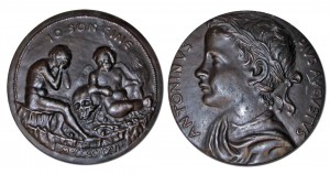
One medal in the collection commemorates the Roman emperor Caracalla. It was struck in Venice, Italy, in 1466. The design is loosely copied from a coin and is attributed to the Venetian artist, Giovanni Boldù, who was active from 1454-1477. It is a large medal (90 mm in diameter) struck in bronze. The obverse has an image of the Roman emperor Caracalla as a boy done in high relief. The youth’s bust facing left is circled by the name ANTONINVS PIVS AVGVSTVS (Antoninus Pius Augustus). The reverse side of the metal pictures the artist seated naked with his head in his hands; before him there is the Genius of Death, resting on a skull; above him, the inscription IO SON FINE; and below him, the date MCCCCLXVI (1466).
As commissioner of a large public bath house (thermae) in Rome, Caracalla fits the pattern of men accomplished in the field of medicine. The building that housed the bath house was constructed during his reign between 212 and 216 AD. It was heated by the ancient hypocast system of under-floor heating, and supplied with water by the Aqua Marcia Aqueduct. The bath house consisted of cold and hot rooms, a pool, gyms, and two libraries. It was free, open to the public and in use until the 6th century, when the hydraulic installations were destroyed. While in use, it provided a place where Romans could bathe and possibly receive health benefits from the baths. Today the ruins of the Baths of Caracalla are a tourist attraction in Rome.
This medal can be viewed in the Rare Book Room by appointment.
~ Gosia Fort
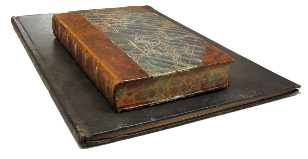
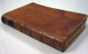
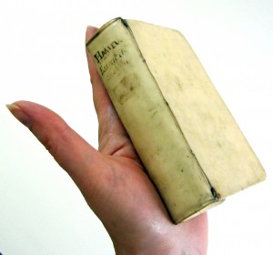 The Falk Library collection includes a pocket-sized edition of this famous work published in the Netherlands in 1648. The book is only 12 cm tall and easily fits in the palm of your hand. It is bound in plain white vellum and has a brief handwritten title on the spine.
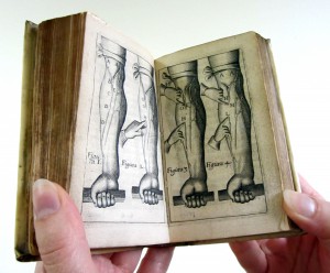 Vellum is best stored in a stable environment with controlled temperature and humidity, such as that in Falk Library’s Rare Book Room.
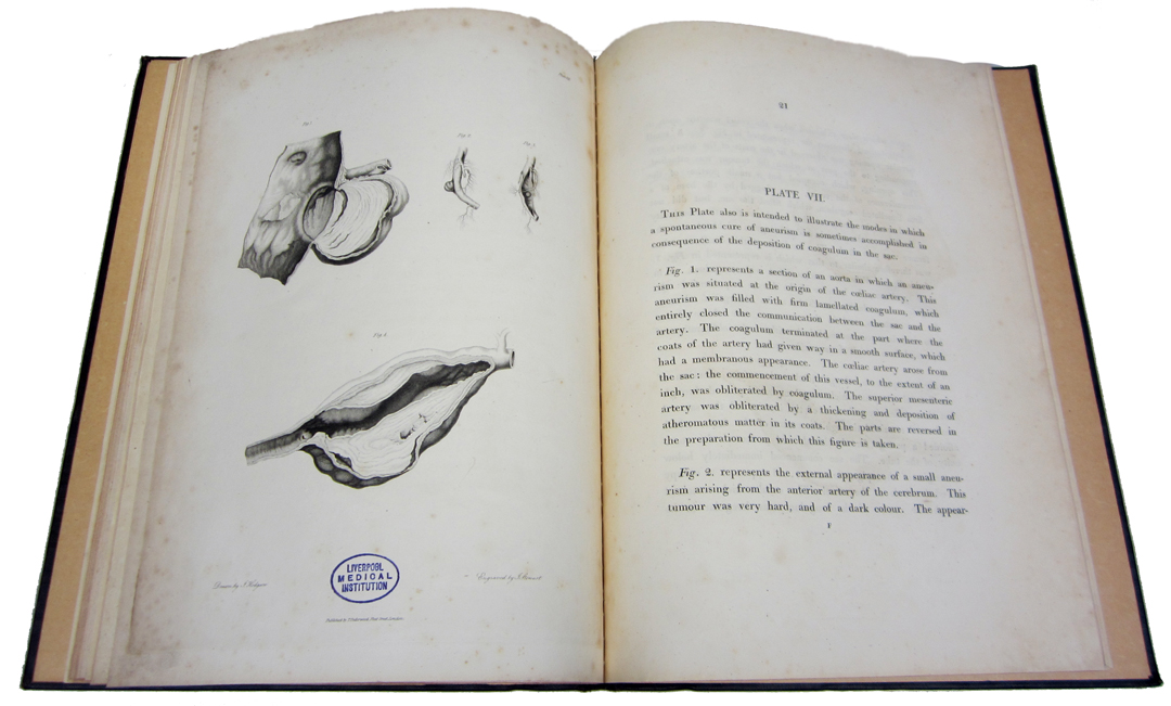
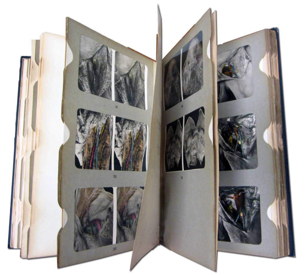 Stereoscopy is a technique for creating depth in an image by presenting two offset images separately to the left and right eye of the viewer. In 1838, Charles Wheatstone showed that the illusion of depth can be created from flat pictures that differed only in horizontal disparity. Stereoscopy became popular after 1849 with the invention of the prism stereoscope by David Brewster. Photography popularized stereograms (side-by-side pictures) even more. Since objects could be experienced in 3-D, stereoscopic images became widely used in books about geography, history, and medicine, among other subjects.
 The original English translation of van Swieten’s work on the hygiene of troops and diseases impacting the military was published in London in 1762, and the first American edition printed by Robert Bell appeared in Philadelphia 14 years later. Thus, the information derived from foreign experiences in military hygiene was made available to young surgeons in the War of Independence. Swieten’s text was enhanced by the addition of several chapters, including “Some Brief Directions to Be Observed by Sea Surgeons in Engagements” by William Northcote (extracted from The Marine Practice of Physic and Surgery, 1770) and, “The Nature and Treatment of Gun-Shot Wounds” by John Ranby (extracted from The Method of Treating Gunshot Wounds, 1744).
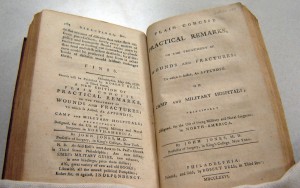 The book, The Diseases Incident to Armies Including Plain Concise Practical Remarks on the Treatment of Wound and Fractures, comes from the collection of our notable donor, the late Dr. Mark M. Ravitch, and is located in the
 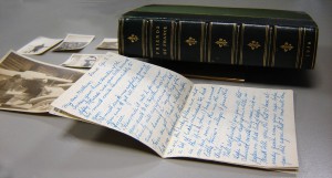 Friends of France: The Field Service of the American Ambulance Described by Its Members. Boston: Houghton Mifflin, 1916.
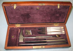 The instruments were produced by Hermann Hernstein & Son. There are three key factors that identify the set as belonging to the Civil War era: (1) the mark of H. Hernstein & Son, which was used from 1862 to 1865, when the company was selling directly to the military under contract; (2) Herstein & Son’s address from 1855 to 1867, 393 Broadway in New York City, is engraved on the instruments; and (3)
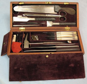 the wooden case contains a single sliding latch not found on any civilian instrument sets of the period.
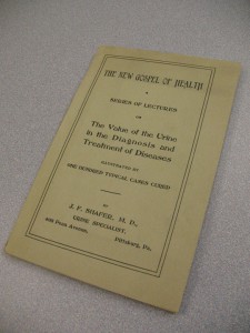 The book includes descriptions of 100 cases illustrating Shafer’s use of urine analysis in the diagnosis and treatment of many illnesses. The catalog of cured ailments ranges from gall stones and jaundice, to ailments such as “tobacco heart.” He assembled these cases with one goal in mind: to share tributes from his patients about the benefits of his diagnostic method. He claimed that the only measurable proof of physician success is a cured patient willing to give testimony about a doctor’s effectiveness. Long before HIPAA regulations protecting patient privacy, Shafer managed to secure his patients’ permission to publish their testimonials to prove his claim. Therefore, all the described cases include detailed observations leading to diagnosis and treatment, and the patients’ testimonials with full names and addresses, so they could be contacted to verify the printed word.