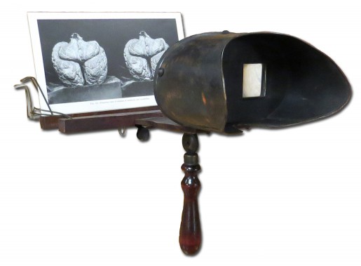
Stereoscopy is a technique that creates the illusion of a 3D image. It is based on a simple principle: when viewing two nearly identical images side by side through prismatic lenses, the eyes blend the two views into one, which is then perceived in three dimensions. Stereoscopic images became widely popular with photography from about 1850 to 1920. These images were a form of entertainment, and even today this technique is used to enhance the teaching power of photography. Falk Library owns several newer anatomy and pathology atlases with stereoscopic illustrations that include their own viewers: Hirsch’s Neuroanatomy (1999) with 3D glasses; Schuknecht’s Stereoscopic Atlas of Mastoidotympanoplastic Surgery (1966) with a folded compact stereo viewer; Bassett’s A Stereoscopic Atlas of Human Anatomy (1965); and Gass’s Stereoscopic Atlas of Macular Diseases (1987, 1997) with a standard reel view-master.
The early stereoscopes and viewers are collectibles. Before the invention of photography, the first stereoscope for viewing drawings was introduced in 1833 by Charles Wheatstone. Later, Oliver Wendell Holmes designed the first handheld viewer which was produced and improved by Joseph Bates.

Falk Library Rare Books Collection
Over the years many other inventors perfected stereoscopes. Hawley C. White was one of them. His company was the largest producer of stereoscopes in the world. He won a prestigious prize at the 1900 International Exhibition in Paris. Many of his 20th century viewers can be identified by the emblem referencing this event. The stereoscope in our collection has his Award Medal depicted in the center of the hood along with the H.C. White name. One of the three card holders has a clasp designed by Truman W. Ingersoll in 1904, thus making the stereoscope traceable to ca.1905. The other two card holders were added later and do not belong to the original viewer. This stereoscope works well with these stereoscopic atlases owned by Falk Library: Cunningham’s Stereoscopic Studies of Anatomy for the Internist (1900); Enderlen and Gasser’s Stereoskopbilder zur Lehre von den Hernien (1906), as described in the February 2012 HSLS Update article, “Treasures from the Rare Book Room: Photography and Medical Books, Part 3”; Kelly’s Dr. J. A. Bodine’s Operation for Inguinal Hernia (1909); Cunningham’s Stereoscopic Studies of Anatomy (ca. 1909); and Jones’s Equilibrium and Vertigo (1918).
These materials can be viewed in the Rare Book Room by appointment.
~ Gosia Fort